
Infectious Bovine Rhinotracheitis
A Major Cause of Viral Abortion in the World
5-60% abortions in non vaccinated herds
The IBR virus is widespread, causes latent infections and can recrudesce, so any cow with a positive IBR titre is a possible carrier. The virus is carried to the placenta, causes placentitis, infects the fetus and kills it in 24 hours. Abortion can happen at any time but usually occurs from 4 months to full-term.
Abortion is not the only reproductive inefficiency associated with this virus.
IBR can directly impact ovarian structures, causing severe lesions on the corpus luteum. This makes developing follicles susceptible to degeneration. These follicles, if able to ovulate, have the potential to release oocytes of reduced viability.
Bovine herpesvirus 1 (BHV-1) is associated with several diseases in cattle:
- Infectious bovine rhinotracheitis (IBR)
- Infectious pustular vulvovaginitis (IPV)
- Balanoposthitis
- Conjunctivitis
- Abortion
- Encephalomyelitis
- Mastitis
Only a single serotype of BHV-1 is recognized, but there are three subtypes of BHV-1:
- BHV-1.1 (respiratory subtype)
- BHV-1.2 (genital subtype)
- BHV-1.3 (encephalitic subtype), now BHV-5
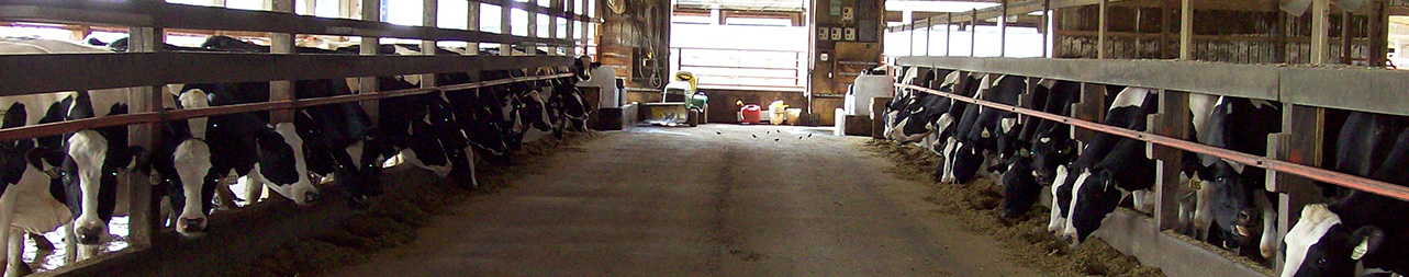
In feedlot cattle, the respiratory form is most common. The viral infection alone is not life-threatening, but predisposes to secondary bacterial pneumonia, which may result in death.
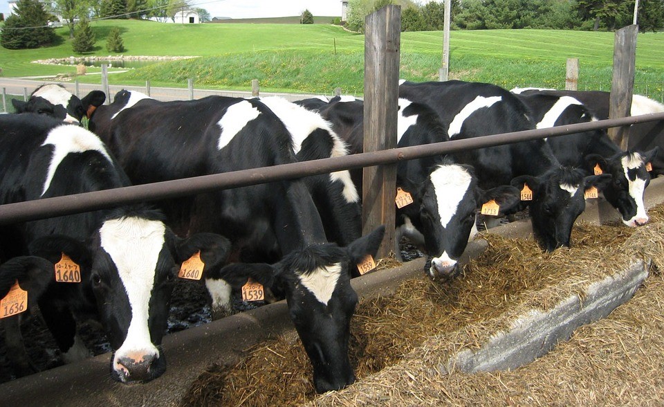
In breeding cattle, abortion or genital infections are more common: Infectious Pustular Balanoposthitis in bulls and Infectious Pustular Vulvovaginitis in cows, within one to three days of mating or close contact with an infected animal.
Transmission can occur:
- Through cows showing no clinical signs or visible lesions that serve as a source of infection for other susceptible animals
- Through artificial insemination with semen from sub-clinically infected bulls
The incubation period for the respiratory and genital forms is two to six days. In the respiratory form, clinical signs range from mild to severe, depending on the presence of secondary bacterial pneumonia.
Respiratory Form
- High fever
- Anorexia
- Coughing
- Excessive salivation
- Nasal discharge that progresses from serous to mucopurulent
- Conjunctivitis with lacrimal discharge
- Inflamed nares (hence the common name “red nose”)
- Dyspnea if the larynx becomes occluded with purulent material
- Clusters of grayish necrotic foci on the mucous membrane of the septal mucosa, just visible inside the external nares; they may later be accompanied by pseudodiphtheritic yellowish plaques
Abortions may occur concurrently with respiratory disease but also may be seen up to 100 days after infection. They can occur regardless of the severity of disease in the dam. Abortions generally occur during the second half of pregnancy, but early embryonic death is possible.
Genital Form
- Frequent urination
- Tailhead elevation
- Mild vaginal discharge
- Swollen vulva, with small papules, erosions and ulcers
- With bacterial infection, there may be inflammation of the uterus and transient infertility, with purulent vaginal discharge for several weeks
- In bulls, similar lesions occur on the penis and prepuce
BHV-1 infection can be severe in young calves and can cause generalized disease. Pyrexia, ocular and nasal discharges, respiratory distress, diarrhea, incoordination, and eventually convulsions and death may occur in a short period after generalized viral infection.
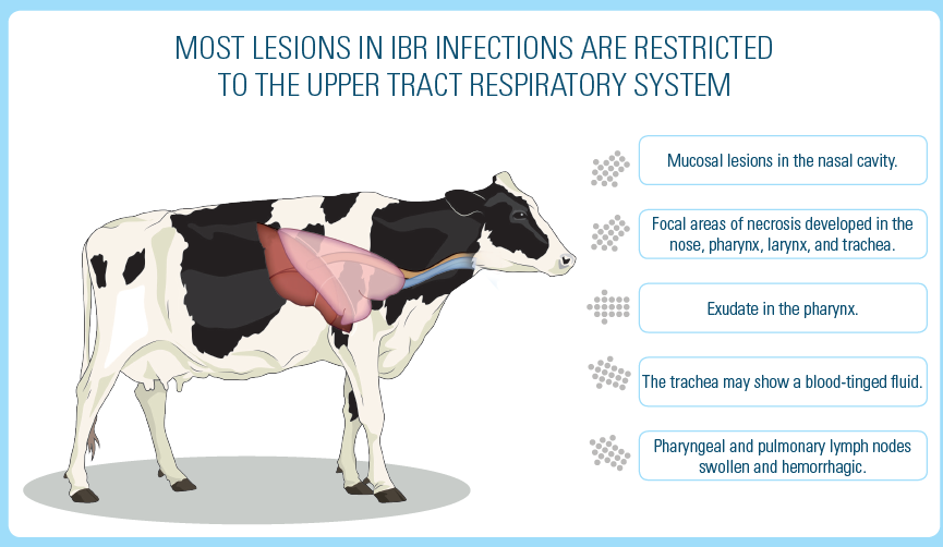
Lesions
In uncomplicated IBR infections, most lesions are in the upper respiratory area.
The viral lesions are often masked by secondary bacterial infections. In young animals with generalized BHV-1 infection, erosions and ulcers overlaid with debris may be found in the nose, esophagus and forestomachs.
White foci may be found in the liver, kidney, spleen and lymph nodes. Aborted fetuses may have pale, focal, necrotic lesions in all tissues, which are especially visible in the liver.
Uncomplicated BHV-1 infections can be diagnosed based on the characteristic signs and lesions. However, because the severity of disease can vary, it is best to differentiate BHV-1 from other viral infections by viral isolation.
- Serology for latent infections
- PCR directly detecting the virus or fluorescent antibody tests on ocular or nasal secretions, for active infections
Antimicrobial therapy is indicated to prevent or treat secondary bacterial pneumonia.
Immunization with modified-live or inactivated virus vaccines generally provides adequate protection against clinical disease.
Biosecurity measures to reduce on-farm risk:
- Diagnose and treat sick animals promptly
- Isolate/remove sick animals from the group or herd
- Keep different age groups and management groups separate
- Reduce direct disease spread from animals
- Take proactive risk reduction measures for farm visitors
Eradication of the virus is possible by a combination of serologic
surveillance, culling of reactors, biosecurity and vaccination.